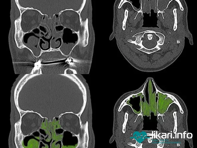
МРТ придаткових пазух носа | MRI of the Paranasal Sinuses
- Категорія: Корисна інформація;
МРТ придаткових пазух носа є сучасним способом діагностики змін структури тканин на ранніх етапах. У процесі дослідження лікар отримує чіткі знімки у тривимірному вигляді, які легко переглядати у різних площинах зі збільшенням зображення. МРТ носових пазух — рідкісне дослідження, але при деяких захворюваннях є необхідним.
МРТ носа роблять, щоб виявити відхилення в порожнинах кісток черепа, ґратчастих лабіринтах, лобових та клиноподібних пазухах, гайморових перегородках, які повідомляються з носом та верхньою частиною щелепи.
Медичні показання до процедури
МРТ носа та приносових пазух роблять за показаннями лікаря. Часто пацієнти затягують із зверненням до фахівця, що може спричинити небажані наслідки. Коли лікар підозрює злоякісне утворення, вдаються до дослідження із контрастом. Для цього спеціальна речовина вводиться у вену та поширюється з кров'ю по всьому організму.
Для того, щоб не пропустити хворобу на ранніх її етапах, необхідно вчасно зробити МРТ носових пазух. До томографії вдаються за таких симптомів:
- часті риніти та втрата нюху;
- аномалії будови черепа в носовій ділянці;
- утруднене дихання;
- запалення придаткових пазух;
- підозри на наявність новоутворень;
- регулярні кровотечі із носа.
Протипоказання щодо МРТ придаткових пазух носа
Це дослідження має низку обмежень, що не дозволяють провести цю діагностику:
- наявність металевих предметів у тілі;
- вагітність (1-й триместр);
- захворювання, що не дозволяють перебувати тілу деякий час у стані нерухомості (хвороба Паркінсона);
- наявність кардіостимулятора, слухового апарату чи інсулінової помпи.
Особливості підготовки до МРТ пазух носа
Діагностика здійснюється у звичайному порядку і не вимагає певних приготувань. Пацієнту слід зняти з себе всі металеві прикраси та предмети.
МРТ носа дитині також проводять, якщо той може деякий час вести себе спокійно та нерухомо.
MRI of the Paranasal Sinuses
MRI of the paranasal sinuses is a modern method for diagnosing structural tissue changes at early stages. During the scan, the doctor obtains clear three-dimensional images that can be viewed in different planes with zoom capability. Although MRI of the sinuses is relatively rare, it is necessary in certain cases.
This procedure is used to detect abnormalities in the skull bone cavities, ethmoidal labyrinths, frontal and sphenoidal sinuses, as well as in the maxillary sinus septa that are connected to the nasal cavity and upper jaw.
Medical Indications for the Procedure
MRI of the nose and paranasal sinuses is performed based on a doctor's referral. Patients often delay seeking help, which can lead to complications. When a malignant tumor is suspected, contrast-enhanced MRI is used. A special substance is injected into a vein and spreads through the bloodstream across the body.
To avoid missing a disease in its early stages, it is important to undergo an MRI scan of the sinuses on time. The procedure is recommended for the following symptoms:
- frequent rhinitis and loss of smell;
- anatomical anomalies in the nasal area of the skull;
- difficulty breathing;
- inflammation of the paranasal sinuses;
- suspected tumors;
- recurring nosebleeds.
Contraindications for MRI of the Paranasal Sinuses
This diagnostic method has certain limitations that may prevent the procedure from being performed:
- presence of metal objects in the body;
- pregnancy (first trimester);
- conditions that prevent the patient from staying still (e.g., Parkinson’s disease);
- presence of a pacemaker, hearing aid, or insulin pump.
Preparation for MRI of the Sinuses
The procedure is usually performed in a standard manner and does not require special preparation. The patient must remove all metal jewelry and objects.
MRI of the nose can also be performed on a child, provided the child can remain calm and still for the duration of the scan.

